المدة الزمنية 6:56
Formation of Urine - Nephron Function, Animation.
تم نشره في 2020/10/08
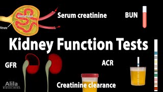
(USMLE topics) Renal physiology - The 3 stages of urine formation. With explanation of the counter current mechanism. Purchase PDF (script of this video + images) here: https://www.alilamedicalmedia.com/-/galleries/pdf-video-scripts-with-images/a-p-basics/-/medias/fba3e922-a451-40a2-8157-140bc5e6a018-formation-of-urine-4-pages-8-images This video is available for instant download licensing here: https://www.alilamedicalmedia.com/-/galleries/all-animations/urinary-system-videos/-/medias/37f5a014-09d2-438b-8053-ec174bd4749f-formation-of-urine-narrated-animation ©Alila Medical Media. All rights reserved. Support us on Patreon and get FREE downloads and other great rewards: patreon.com/AlilaMedicalMedia The kidneys filter blood plasma, removing metabolic wastes, toxins from the body and excrete them in the form of urine. During this process, they also maintain constant volume and composition of the blood, or homeostasis. Blood enters the kidney via the renal artery, which divides to smaller arteries and finally arterioles. The arterioles get into contact with functional units of the kidney called nephrons. This is where blood filtration and urine formation take place. The filtered blood is then collected in to a series of larger veins and exits the kidney through the renal vein. The urine is collected in collecting ducts and leaves the kidney via the ureters. A nephron consists of 2 major parts: Bowman’s capsule; and a long renal tubule. Renal tubules of several nephrons connect to a common collecting duct. There are 3 steps in the formation of urine: glomerular filtration, tubular reabsorption and secretion, water conservation. Blood enters the Bowman’s capsule via the afferent arteriole, passes through a ball of capillaries called the glomerulus, and leaves via the efferent arteriole. Hydrostatic and osmotic pressures drive water and solutes from blood plasma through a filtration membrane into the capsular space of nephron. These include water, inorganic ions, glucose, amino acids and various metabolic wastes such as urea and creatinine. The amount of filtrate produced per minute is called glomerular filtration rate, or GFR. The GFR is kept at a stable value by several feedback mechanisms within the kidneys. This is known as renal autoregulation. The GFR is also under sympathetic and hormonal control. The proximal convoluted tubule, reabsorbs about two thirds of the filtrate. In this process, water and solutes are driven through the epithelial cells that line the tubule into the extracellular space. They are then taken up by the peritubular capillaries. Sodium re-absorption is most important, as it creates osmotic pressure that drives water and electrical gradient that drives negatively charged ions. Sodium level inside the epithelial cells is kept low thanks to the sodium-potassium pumps that constantly pump sodium ions out into the extracellular space. This creates a concentration gradient that favors sodium diffusion from tubular fluid into the cells. Sodium is absorbed by symport proteins that also bind glucose and some other solutes. About half of nitrogenous wastes also re-absorbs back to the bloodstream. Some of the re-absorption also occurs by the paracellular route through tight junctions between the epithelial cells. At the same time, tubular secretion also takes place. The main function of the loop of Henle is to create and maintain an osmolarity gradient in the medulla that enables the collecting ducts to concentrate urine at a later stage. The ascending limb of the loop actively pumps sodium out making the medulla “salty”. The descending limp of the loop is permeable to water but much less to sodium. As the water exits the tubule by osmosis, the filtrate gets more and more concentrated as it reaches the bottom. The ascending limb, on the other hand, is permeable to ions but not water. As a result, the filtrate loses sodium as it goes up and becomes more diluted at the top of the loop. Re-absorption and secretion in the distal convoluted tubule are under control of various hormones. The main function of the collecting duct is to concentrate urine and therefore conserve water. As it gets saltier deep in the medulla, the filtrate loses more and more water as it flows down the collecting duct. The collecting duct is also under hormonal control so it can adjust the amount of re-absorbed water accordingly to the body’s state of hydration. All images/videos by Alila Medical Media are for information purposes ONLY and are NOT intended to replace professional medical advice, diagnosis or treatment. Always seek the advice of a qualified healthcare provider with any questions you may have regarding a medical condition.
الفئة
عرض المزيد


تعليقات - 290
مقاطع الفيديو ذات الصلة على Formation of Urine - Nephron Function, Animation.: